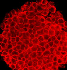
P19细胞

P19是一种源自小鼠胚胎生殖细胞瘤的胚胎性癌细胞系,可以分化为三个胚层的细胞类型,并且是最具特征性的胚胎性癌细胞系,可以通过不同的特异性治疗方法,诱导为心肌细胞和神经元细胞,例如将密集的P19细胞暴露于二甲基亚砜(DMSO)时会诱导其分化为心肌和骨骼肌细胞,而将P19细胞暴露于视黄酸(RA)时,则可以将其分化为神经元细胞[1]。
起源
[编辑]如果具侵袭性的癌细胞增殖并转移到其他部位,可能导致患者死亡,然而研究人员也能利用这些细胞来研究癌细胞的发育过程,以便找到更具体的治疗方法。对于生物学家来说,起源于生殖细胞瘤的胚胎性癌细胞系是进行癌细胞发育研究的良好对象。1982年,有科研人员将仅7.5天大的小鼠胚胎移植到睾丸中,以诱导肿瘤的生长。从具有整倍体核型的原发性肿瘤中,分离出含有未分化干细胞的细胞培养物。这些干细胞被称为P19细胞[2]。这些衍生的P19细胞在没有饲养细胞(feeder cells)存在的情况下迅速生长,并且易于维护。通过将P19细胞注射到另一个小鼠品系的胚泡中,可以确认P19细胞的多能性。在身为受体的小鼠中,三个胚层都有组织生长[3]。基于后来的研究,科研人员进一步从原始的P19细胞衍生了四个亚型,分别为P19S18、P19D3、P19RAC65及P19C16。这些亚型之间的不同之处是响应于RA或DMSO处理而分化为神经元细胞或肌肉细胞的能力[3][4][5]。由于P19细胞具有多能性,因此这些衍生的细胞系可以是外胚层、中胚层及内胚层样细胞[6]。
细胞分化
[编辑]由于P19细胞具有稳定的染色体组成,所以其可以保持着指数的增长。因为胚胎癌可以分化成三个胚层的细胞,所以P19细胞也可以分化成外胚层、中胚层和内胚层样细胞。当胚胎性癌细胞进行高密度的培养时,它们会开始分化[7]。通过将细胞聚集到胚胎的体内,EC细胞可以进行分化[8]。在密集的类胚体细胞中,添加对细胞无毒性的浓度的药物可以诱导P19细胞分化为特定的细胞系[1]。两种最常见和最有效的药物是RA和DMSO。有研究表明一定浓度的RA也可以诱导P19细胞分化为神经元细胞,其中包括神经元和神经胶质细胞[9],而0.5%至1%的DMSO则导致P19细胞分化为心肌或骨骼肌细胞。在RA治疗方法中,聚集后可以鉴定神经元、星形胶质细胞及成纤维细胞。已进行分化的细胞也具有胆碱乙酰转移酶和乙酰胆碱酯酶活性[10]。当用DMSO处理细胞后5天后时,已有心肌细胞发育,而8天后则出现骨骼肌细胞。这些研究表明,暴露于药物会导致多能性的P19细胞分化为不同的细胞层。由于RA或DMSO的浓度对细胞并没有毒性,因此药物特异性分化是由于细胞被诱导的缘故。目前已经产生P19细胞的突变体,以研究药物特异性分化的机制[10]。此外,通过研究基因表达或产生P19细胞突变体,可以研究与神经发生及肌肉生成有关的信号通路。
神经发生
[编辑]利用RA处理未分化的P19细胞,可以特异性诱导它们成为神经元细胞。使用1μM至3μM 剂量的RA可以令神经元成为细胞内含量最丰富的细胞类型[4]。经过有关处理的神经元在六天至九天内成为细胞内最高含量的种群,而神经纤维丝蛋白、HNK-1抗原和破伤风毒素结合位点等神经元标记物会以最高的水平表达[11]。经过六至九天的治疗,神经元数量会下降,原因可能是由于非神经元细胞比神经元细胞更快速地增殖。暴露于RA10天后,可以使用神经胶质纤维酸性蛋白检测星形胶质细胞,因为该蛋白是神经胶质细胞的特异性标志物。除了分化成为神经元和星形胶质细胞外,P19细胞还可以分化为寡突胶质细胞,并且可以使用特异性标记物、髓鞘相关糖蛋白及CNPase进行检测。当经RA诱导的细胞移植到大脑中时,寡突胶质细胞也会发育并迁移至成纤维束[12]。
维生素A酸不仅可以诱导P19细胞,还可以诱导着其他祖细胞或胚胎干细胞的分化。由于经RA处理后的细胞,不会立即表达神经元标记基因,因此RA必须启动某种途径来进行细胞分化。许多学者使用P19细胞研究RA诱导的机制,包括产生RA受体基因的突变等位基因,以及研究暴露于RA时,受体基因、同源异形基因及视黄醇结合蛋白质的表达[13][14]。这些研究表明,P19细胞可用于研究干扰特定细胞途径的药物机制,故而是有关领域上良好的体外模型系统。
此外,通过利用RA诱导的P19细胞神经发生的能力,许多研究人员开始确定神经或神经胶质发生的体外分化机制。使用基因表达或产生相关基因的等位基因研究了几种相关信号通路,包括Wnt/β-catenin信号通路、Notch信号通路,以及刺猬信号通路[15][16][17]。
肌肉生成
[编辑]与视黄酸相同,DMSO诱导的分化不是P19细胞特有的。它也可以诱导神经母细胞瘤细胞、肺癌细胞和小鼠ES细胞[18][19][20]。在浓度为0.5%至1%的DMSO中,DMSO诱导P19细胞聚集并处理中胚层和内胚层细胞类型。聚集和分化过程中的细胞机制仍未被完全研究。然而有研究表明,细胞的信息传递在P19细胞的肌肉分化中起著重要作用,或可以解释细胞需要先聚集才能进行肌肉分化的原因。为了阐明P19细胞中肌发生的机制,有研究人员发现GATA-4、MEF2c、Msx-1、Nkx2.5、MHox、Msx-2及MLP等心脏特异性转录因子在分化过程中发生变化[6],例如在细胞经DMSO诱导后,GATA-4、NKx2.5和MEF2c的表达均被上调[21][22]。P19细胞还用于研究心脏的分化和肌肉生成的机制。骨塑型蛋白(BMP)信号通路是在P19细胞中研究最深入的信号通路。它们通过产生过度表达BMP拮抗剂的P19CL6noggin细胞,发现其经用1%DMSO处理后,突变的细胞不会分化为心肌细胞,这表明BMPs在该系统中对于心肌细胞的分化是必不可少的东西,有关证据表明TAK1、Nkx-2.5及GATA-4在心源性BMP信号通路中十分重要[23]。
未来研究方向
[编辑]P19细胞可以在体外形成神经元细胞和肌肉细胞。P19细胞与其他胚胎干细胞相比,更容进行易维护和培养,因此它们是在体外进行发育研究的便捷模型。 操作该细胞系以表达或敲除某些基因的技术,可以用于详细研究信号传导途径,以及肌肉生成和神经生成的蛋白质表达调控。经扩展的研究还可以阐明心脏或大脑发育及成熟的后期阶段。
参考资料
[编辑]- ^ 1.0 1.1 McBurney, MW; Rogers, BJ. Isolation of male embryonal carcinoma cells and their chromosome replication patterns.. Developmental biology. 1982-02, 89 (2): 503–8 [2020-01-08]. PMID 7056443. doi:10.1016/0012-1606(82)90338-4.[永久失效链接]
- ^ McBurney, MW. P19 embryonal carcinoma cells.. The International journal of developmental biology. 1993-03, 37 (1): 135–40 [2020-01-08]. PMID 8507558.[永久失效链接]
- ^ 3.0 3.1 Rossant, J; McBurney, MW. The developmental potential of a euploid male teratocarcinoma cell line after blastocyst injection.. Journal of embryology and experimental morphology. 1982-08, 70: 99–112 [2020-01-08]. PMID 7142904.[永久失效链接]
- ^ 4.0 4.1 Fahnestock, M; Koshland DE, Jr. Control of the receptor for galactose taxis in Salmonella typhimurium.. Journal of bacteriology. 1979-02, 137 (2): 758–63 [2020-01-08]. PMID 370099.[永久失效链接]
- ^ Craine, BL; Rupert, CS. Deoxyribonucleic acid-membrane interactions near the origin of replication and initiation of deoxyribonucleic acid synthesis in Escherichia coli.. Journal of bacteriology. 1979-02, 137 (2): 740–5 [2020-01-08]. PMID 370098.[永久失效链接]
- ^ 6.0 6.1 van der Heyden, MA; Defize, LH. Twenty one years of P19 cells: what an embryonal carcinoma cell line taught us about cardiomyocyte differentiation.. Cardiovascular research. 2003-05-01, 58 (2): 292–302 [2020-01-08]. PMID 12757864. doi:10.1016/s0008-6363(02)00771-x.[永久失效链接]
- ^ McBurney, MW. Clonal lines of teratocarcinoma cells in vitro: differentiation and cytogenetic characteristics.. Journal of cellular physiology. 1976-11, 89 (3): 441–55 [2020-01-08]. PMID 988033. doi:10.1002/jcp.1040890310.[永久失效链接]
- ^ Martin, GR; Evans MJ. Multiple differentiation of clonal teratocarcinoma stem cells following embryoid body formation in vitro. Cell. 1975, 6 (4): 467–74. doi:10.1016/0092-8674(75)90035-5.
- ^ Edwards, MK; Harris, JF; McBurney, MW. Induced muscle differentiation in an embryonal carcinoma cell line.. Molecular and cellular biology. 1983-12, 3 (12): 2280–6 [2020-01-08]. PMID 6656767. doi:10.1128/mcb.3.12.2280.[永久失效链接]
- ^ 10.0 10.1 Jones-Villeneuve, E M; Rudnicki, M A; Harris, J F; McBurney, M W. Retinoic acid-induced neural differentiation of embryonal carcinoma cells.. Molecular and Cellular Biology. 1983-12, 3 (12): 2271–2279 [2020-01-08]. PMID 6656766. doi:10.1128/mcb.3.12.2271.[永久失效链接]
- ^ McBurney, MW; Reuhl, KR; Ally, AI; Nasipuri, S; Bell, JC; Craig, J. Differentiation and maturation of embryonal carcinoma-derived neurons in cell culture.. The Journal of neuroscience : the official journal of the Society for Neuroscience. 1988-03, 8 (3): 1063–73 [2020-01-08]. PMID 2894413.[永久失效链接]
- ^ Staines, WA; Craig, J; Reuhl, K; McBurney, MW. Retinoic acid treated P19 embryonal carcinoma cells differentiate into oligodendrocytes capable of myelination.. Neuroscience. 1996-04, 71 (3): 845–53 [2020-01-08]. PMID 8867053. doi:10.1016/0306-4522(95)00494-7.[永久失效链接]
- ^ Pratt, MA; Kralova, J; McBurney, MW. A dominant negative mutation of the alpha retinoic acid receptor gene in a retinoic acid-nonresponsive embryonal carcinoma cell.. Molecular and cellular biology. 1990-12, 10 (12): 6445–53 [2020-01-08]. PMID 2174108. doi:10.1128/mcb.10.12.6445.[永久失效链接]
- ^ Chen, Y; Reese, DH. The retinol signaling pathway in mouse pluripotent P19 cells.. Journal of cellular biochemistry. 2011-10, 112 (10): 2865–72 [2020-01-08]. PMID 21618588. doi:10.1002/jcb.23200.[永久失效链接]
- ^ Nye, JS; Kopan, R; Axel, R. An activated Notch suppresses neurogenesis and myogenesis but not gliogenesis in mammalian cells.. Development (Cambridge, England). 1994-09, 120 (9): 2421–30 [2020-01-08]. PMID 7956822.[永久失效链接]
- ^ Hamada-Kanazawa, M; Ishikawa, K; Nomoto, K; Uozumi, T; Kawai, Y; Narahara, M; Miyake, M. Sox6 overexpression causes cellular aggregation and the neuronal differentiation of P19 embryonic carcinoma cells in the absence of retinoic acid.. FEBS letters. 2004-02-27, 560 (1-3): 192–8 [2020-01-08]. PMID 14988021. doi:10.1016/S0014-5793(04)00086-9.[永久失效链接]
- ^ Tan, Y; Xie, Z; Ding, M; Wang, Z; Yu, Q; Meng, L; Zhu, H; Huang, X; Yu, L; Meng, X; Chen, Y. Increased levels of FoxA1 transcription factor in pluripotent P19 embryonal carcinoma cells stimulate neural differentiation.. Stem cells and development. 2010-09, 19 (9): 1365–74 [2020-01-08]. PMID 19916800. doi:10.1089/scd.2009.0386.[永久失效链接]
- ^ Lako, M; Lindsay, S; Lincoln, J; Cairns, PM; Armstrong, L; Hole, N. Characterisation of Wnt gene expression during the differentiation of murine embryonic stem cells in vitro: role of Wnt3 in enhancing haematopoietic differentiation.. Mechanisms of development. 2001-05, 103 (1-2): 49–59 [2020-01-09]. PMID 11335111. doi:10.1016/s0925-4773(01)00331-8.[永久失效链接]
- ^ Tralka, TS; Rabson, AS. Cilia formation in cultures of human lung cancer cells treated with dimethyl sulfoxide.. Journal of the National Cancer Institute. 1976-12, 57 (6): 1383–8 [2020-01-09]. PMID 1003564. doi:10.1093/jnci/57.6.1383.[永久失效链接]
- ^ Uz, Littauer; C, Palfrey; Y, Kimhi; I, Spector. Induction of Differentiation in Mouse Neuroblastoma Cells. National Cancer Institute monograph. 1978-05 [2020-01-09]. PMID 748753 (英语).[永久失效链接]
- ^ Skerjanc, IS; Petropoulos, H; Ridgeway, AG; Wilton, S. Myocyte enhancer factor 2C and Nkx2-5 up-regulate each other's expression and initiate cardiomyogenesis in P19 cells.. The Journal of biological chemistry. 1998-12-25, 273 (52): 34904–10 [2020-01-09]. PMID 9857019. doi:10.1074/jbc.273.52.34904.[永久失效链接]
- ^ Grépin, C; Nemer, G; Nemer, M. Enhanced cardiogenesis in embryonic stem cells overexpressing the GATA-4 transcription factor.. Development (Cambridge, England). 1997-06, 124 (12): 2387–95 [2020-01-09]. PMID 9199365.[永久失效链接]
- ^ Monzen, K; Shiojima, I; Hiroi, Y; Kudoh, S; Oka, T; Takimoto, E; Hayashi, D; Hosoda, T; Habara-Ohkubo, A; Nakaoka, T; Fujita, T; Yazaki, Y; Komuro, I. Bone morphogenetic proteins induce cardiomyocyte differentiation through the mitogen-activated protein kinase kinase kinase TAK1 and cardiac transcription factors Csx/Nkx-2.5 and GATA-4.. Molecular and cellular biology. 1999-10, 19 (10): 7096–105 [2020-01-09]. PMID 10490646. doi:10.1128/mcb.19.10.7096.[永久失效链接]
